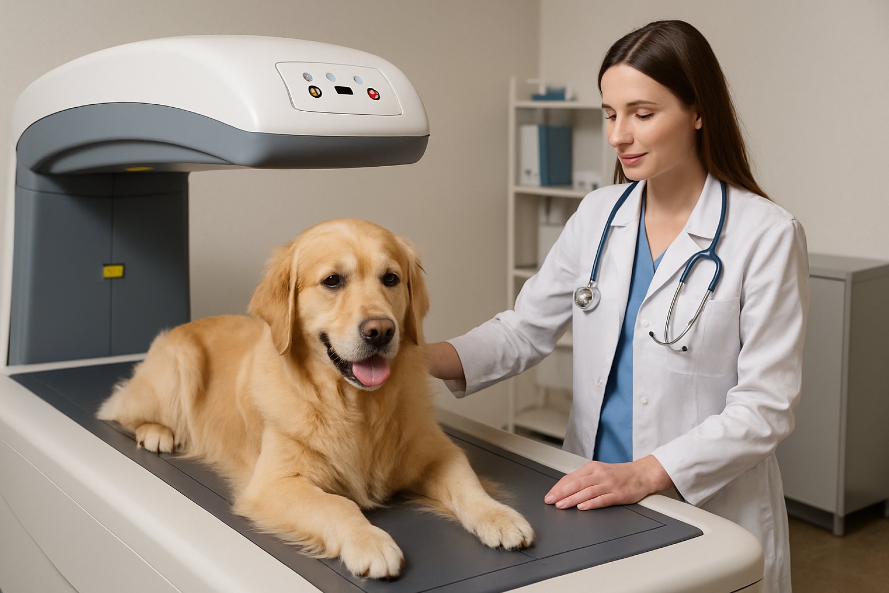
Unlocking the Power of Dual-energy X-ray Absorptiometry (DXA) in Veterinary Medicine: The New Gold Standard for Animal Bone and Body Composition Analysis
- Introduction to DXA Technology in Veterinary Medicine
- How DXA Works: Principles and Mechanisms
- Clinical Applications: Diagnosing Bone Density and Body Composition in Animals
- Benefits of DXA Over Traditional Imaging Methods
- Case Studies: Real-World Impact of DXA in Veterinary Practice
- Limitations and Considerations for Veterinary Use
- Future Directions: Innovations and Research in Veterinary DXA
- Conclusion: The Evolving Role of DXA in Animal Healthcare
- Sources & References
Introduction to DXA Technology in Veterinary Medicine
Dual-energy X-ray Absorptiometry (DXA) is an advanced imaging modality that has become increasingly valuable in veterinary medicine for the quantitative assessment of bone mineral density (BMD) and body composition. Originally developed for human healthcare, DXA technology utilizes two X-ray beams at different energy levels to differentiate between bone and soft tissue, providing precise measurements of bone mass, lean tissue, and fat content. This non-invasive technique is now being adapted for a variety of animal species, offering veterinarians a reliable tool for diagnosing metabolic bone diseases, monitoring osteoporosis, and evaluating the effects of nutritional or pharmacological interventions.
The application of DXA in veterinary practice extends beyond research settings, with growing use in clinical diagnostics and animal health management. Its high accuracy and reproducibility make it particularly useful for longitudinal studies and for tracking changes in bone and body composition over time. Furthermore, DXA’s relatively low radiation dose and rapid scan times are advantageous for animal welfare, minimizing stress and risk during procedures. The technology is also being explored for use in exotic and zoo animals, broadening its impact across diverse veterinary fields.
Despite its benefits, the implementation of DXA in veterinary medicine presents unique challenges, such as the need for species-specific calibration and the adaptation of human-based protocols to accommodate anatomical differences in animals. Ongoing research and technological advancements continue to refine these applications, enhancing the utility and accessibility of DXA for veterinary professionals. For further information on the principles and veterinary applications of DXA, refer to resources from the American Veterinary Medical Association and the American College of Veterinary Radiology.
How DXA Works: Principles and Mechanisms
Dual-energy X-ray Absorptiometry (DXA) operates on the principle of differential attenuation of X-rays by various tissues in the body. The system utilizes two distinct X-ray energy levels, which are directed through the animal’s body. As these X-rays pass through, tissues such as bone, lean muscle, and fat absorb the energy to different extents. The detector on the opposite side of the body measures the amount of X-ray energy that emerges, and sophisticated algorithms use this data to distinguish between bone mineral content, lean tissue mass, and fat mass. This dual-energy approach allows for precise quantification of body composition, as the attenuation coefficients for bone and soft tissues differ significantly at the two energy levels.
In veterinary medicine, DXA scanners are adapted to accommodate a range of animal sizes and species, from small rodents to large companion animals. The animal is typically positioned on a scanning table, and the X-ray source and detector move in a synchronized manner to capture a series of images. The resulting data are processed to generate detailed two-dimensional maps of bone density and soft tissue distribution. Importantly, the radiation dose used in DXA is relatively low, making it a safe option for repeated measurements in both clinical and research settings. The accuracy and reproducibility of DXA have made it a gold standard for assessing bone mineral density and body composition in veterinary patients, supporting both diagnostic and longitudinal studies American Veterinary Medical Association, National Center for Biotechnology Information.
Clinical Applications: Diagnosing Bone Density and Body Composition in Animals
Dual-energy X-ray Absorptiometry (DXA) has become an invaluable tool in veterinary medicine for the clinical assessment of bone density and body composition in a variety of animal species. Its primary application lies in the diagnosis and monitoring of metabolic bone diseases, such as osteoporosis and osteopenia, particularly in companion animals and research models. By providing precise measurements of bone mineral density (BMD), DXA enables veterinarians to detect early bone loss, evaluate fracture risk, and monitor the efficacy of therapeutic interventions. This is especially relevant in aging pets, animals with endocrine disorders, or those receiving long-term corticosteroid therapy, where bone health is a significant concern.
Beyond bone health, DXA is widely used to assess body composition, including the quantification of lean mass, fat mass, and regional fat distribution. This capability is crucial for managing obesity, a growing problem in domestic animals, and for tailoring nutritional and exercise regimens. In performance animals, such as racehorses and working dogs, DXA provides objective data to optimize conditioning and monitor muscle development. The technique’s non-invasive nature and high reproducibility make it suitable for longitudinal studies and repeated measurements, minimizing stress and risk to the animal.
Recent advances have expanded DXA’s use to exotic and zoo animals, enhancing the understanding of species-specific bone and body composition norms. As the technology becomes more accessible, its role in both clinical practice and research is expected to grow, supporting evidence-based veterinary care and improving animal welfare (American Veterinary Medical Association; National Center for Biotechnology Information).
Benefits of DXA Over Traditional Imaging Methods
Dual-energy X-ray Absorptiometry (DXA) offers several significant advantages over traditional imaging modalities such as radiography and computed tomography (CT) in veterinary medicine. One of the primary benefits is its superior accuracy and precision in quantifying bone mineral density (BMD) and body composition, including lean and fat mass. Unlike standard radiographs, which provide only qualitative or semi-quantitative assessments, DXA delivers objective, reproducible measurements that are critical for diagnosing and monitoring metabolic bone diseases, such as osteoporosis, in animals American Veterinary Medical Association.
DXA is also associated with lower radiation exposure compared to CT scans, making it a safer option for both animals and veterinary staff, especially when repeated assessments are necessary. The procedure is non-invasive and typically requires minimal sedation, reducing stress and risk for the patient. Additionally, DXA scans are relatively quick to perform, allowing for efficient throughput in clinical settings American College of Veterinary Radiology.
Another notable advantage is DXA’s ability to differentiate between soft tissue and bone, enabling comprehensive evaluation of body composition. This is particularly valuable in research and clinical practice for monitoring obesity, cachexia, or muscle wasting in companion animals. Furthermore, the standardized protocols and automated analysis provided by modern DXA systems enhance consistency and reduce operator-dependent variability, which is a common limitation in traditional imaging methods World Small Animal Veterinary Association.
Case Studies: Real-World Impact of DXA in Veterinary Practice
Case studies have demonstrated the significant real-world impact of Dual-energy X-ray Absorptiometry (DXA) in veterinary practice, particularly in the diagnosis and management of metabolic bone diseases, obesity, and body composition analysis in companion animals. For instance, a clinical case involving a middle-aged canine with suspected osteopenia utilized DXA to accurately quantify bone mineral density (BMD), leading to a tailored therapeutic regimen and improved patient outcomes. Similarly, DXA has been instrumental in feline medicine, where it enabled early detection of osteoporosis in geriatric cats, allowing for timely intervention and monitoring of treatment efficacy.
In the context of obesity management, DXA has provided veterinarians with precise measurements of fat and lean mass, surpassing the limitations of traditional body condition scoring. A notable case involved a weight management program for a group of overweight dogs, where DXA assessments guided dietary adjustments and exercise plans, resulting in measurable reductions in fat mass and improved overall health. These outcomes underscore the value of DXA in both individual patient care and broader clinical research.
Furthermore, DXA has been applied in exotic and zoo animal medicine, where non-invasive and accurate assessment of skeletal health is critical. For example, DXA was used to monitor bone density in captive reptiles, informing husbandry changes that mitigated metabolic bone disease. Collectively, these case studies highlight the versatility and clinical utility of DXA, supporting its integration into routine veterinary diagnostics and research protocols (American Veterinary Medical Association; National Center for Biotechnology Information).
Limitations and Considerations for Veterinary Use
While Dual-energy X-ray Absorptiometry (DXA) has become an invaluable tool for assessing bone mineral density and body composition in veterinary medicine, several limitations and considerations must be addressed for its effective use in animal patients. One primary limitation is the lack of species-specific reference data. Most DXA systems and normative databases are designed for human use, making it challenging to interpret results accurately for different animal species, breeds, and sizes. This can lead to potential misclassification of bone health or body composition in veterinary patients American Veterinary Medical Association.
Another consideration is the need for patient immobilization. Unlike human patients, animals often require sedation or anesthesia to remain still during the scan, which introduces additional risks and logistical challenges. Furthermore, variations in body conformation, fur thickness, and positioning can affect the accuracy and reproducibility of DXA measurements in animals National Center for Biotechnology Information.
Cost and accessibility also pose significant barriers. DXA equipment is expensive and not widely available in general veterinary practice, often limiting its use to research institutions or specialty clinics. Additionally, the interpretation of DXA results requires specialized training, and there is a need for standardized protocols tailored to veterinary applications American College of Veterinary Radiology.
In summary, while DXA offers valuable insights for veterinary medicine, its application is constrained by technical, logistical, and interpretive challenges that must be carefully considered to ensure accurate and meaningful results.
Future Directions: Innovations and Research in Veterinary DXA
The future of Dual-energy X-ray Absorptiometry (DXA) in veterinary medicine is poised for significant innovation, driven by advances in imaging technology, data analytics, and species-specific research. One promising direction is the development of portable and more affordable DXA units, which could facilitate broader clinical and field use, especially in equine and large animal practice. Enhanced software algorithms are being designed to improve the accuracy of bone mineral density (BMD) and body composition measurements across a wider range of species, addressing current limitations related to anatomical differences and size variability.
Artificial intelligence (AI) and machine learning are increasingly being integrated into DXA analysis, enabling automated interpretation and more precise quantification of skeletal and soft tissue parameters. These technologies may also support the creation of large, species-specific reference databases, improving diagnostic accuracy for conditions such as osteoporosis, obesity, and metabolic bone disease in companion and exotic animals. Additionally, research is underway to adapt DXA for longitudinal monitoring, allowing veterinarians to track changes in bone and muscle health over time and assess the efficacy of therapeutic interventions.
Collaborative efforts between veterinary institutions and human medical imaging companies are fostering the adaptation of advanced DXA protocols, such as three-dimensional imaging and regional analysis, for veterinary applications. As these innovations progress, regulatory guidance and standardized protocols will be essential to ensure consistency and reliability in clinical and research settings. Continued investment in veterinary-specific DXA research is expected to expand its utility, ultimately enhancing animal health and welfare through improved diagnostic and monitoring capabilities (American Veterinary Medical Association).
Conclusion: The Evolving Role of DXA in Animal Healthcare
The application of Dual-energy X-ray Absorptiometry (DXA) in veterinary medicine has evolved significantly, transitioning from a primarily research-based tool to an increasingly valuable asset in clinical practice. As the technology becomes more accessible and its protocols are refined for a variety of animal species, DXA is poised to play a central role in the assessment of bone mineral density, body composition, and metabolic health in companion animals, livestock, and even exotic species. Its non-invasive nature, high precision, and ability to provide detailed quantitative data make it particularly advantageous for monitoring disease progression, evaluating therapeutic interventions, and guiding nutritional management in veterinary patients.
Recent advancements in DXA software and hardware have improved its accuracy and adaptability, allowing for more species-specific calibrations and the development of reference ranges tailored to different breeds and ages. These improvements are fostering greater confidence among veterinary professionals in the clinical utility of DXA, not only for diagnosing conditions such as osteoporosis and obesity but also for supporting preventive healthcare strategies. Furthermore, the integration of DXA data with other diagnostic modalities and electronic health records is enhancing the holistic management of animal health.
Looking ahead, continued research and collaboration between veterinary clinicians, researchers, and industry partners will be essential to fully realize the potential of DXA in animal healthcare. As guidelines and best practices are established, DXA is expected to become a standard component of comprehensive veterinary diagnostics, ultimately contributing to improved animal welfare and clinical outcomes American Veterinary Medical Association American College of Veterinary Radiology.
Sources & References
- American Veterinary Medical Association
- American College of Veterinary Radiology
- National Center for Biotechnology Information
- World Small Animal Veterinary Association


